Deformable image registration (DIR) is useful in many aspects of radiation therapy, including contouring, dose deformation, dose tracking and adaptive planning. DIR functionality is incorporated into the RayStation treatment planning system, which means it is integrated into the planning workflow, without the need to export and transfer data between different programs. Research collaborations with partners such as Princess Margaret Cancer Center in Toronto, Canada, have ensured that RayStation meets customers’ needs in a variety of workflow scenarios. Deformable registration is an important aspect of adaptive therapy and RaySearch has dedicated considerable research and development resources to pioneering adaptive planning. From the outset, RayStation was developed with the infrastructure necessary for managing multiple data sets and modalities with various deformation fields. Currently, two algorithms for deformable image registration (DIR) are available in RayStation, giving the clinician a high degree of flexibility when it comes to managing image modalities and data sets of varying image quality. These are outlined in detail below.
HYBRID DIR
The algorithm for hybrid DIR in RayStation is called ANACONDA (“anatomically constrained deformation algorithm”). ANACONDA combines image information, such as intensities, with anatomical information provided by contoured image sets. This makes it possible to maintain the integrity of anatomical structures, even in areas where image information is less reliable. Algorithms that are purely based on image information are limited as they can only regularize the deformation field by applying some degree of smoothing. The supported image modalities are CT, CBCT and MR.
It has been validated that ANACONDA performs well in comparison with other algorithms and that it gives good results for CT/CT¹ and CT/CBCT DIR⁶. ANACONDA has also been applied in MR/MR DIR (same sequence)⁷.
It is interesting to note that accuracy results for deformable registration can vary significantly between centers because different routines are followed. A recent study found that RayStation gave higher levels of accuracy than the other systems evaluated when effective routines for deformable registration were employed¹. The study also showed good results for default settings and independently confirmed some of the results of a 2015 study by Weistrand and Svensson⁶.
Algorithmic details
ANACONDA is based on a mathematical formulation where the registration problem is formulated as a non-linear optimization problem and addressed with a specifically tailored solver that was developed in-house. The objective function is a weighted sum of three non-linear terms:
Image similarity term – measured through the correlation coefficient
Grid regularization term – designed to keep the deformed image grid smooth and invertible
Penalty term – added to the optimization problem when controlling structures are used, in order to deform the selected structure in the reference image to the corresponding structure in the target image
The weights for the different terms in the objective function have been optimized to perform well on a large set of data, but can be changed through scripting for advanced users.
ANACONDA is GPU-accelerated. It has been implemented on CPU and GPU using the same code base, ensuring identical results regardless of hardware.
RayStation’s algorithms for adaptive therapy and deformable registration work extremely well with our protocolized workflow. When a new plan is proposed, a radiation therapist will assess the new GTV and CTV, and we do most corrections using the 3D mesh tool. It takes just a few drag and drop moves. The algorithm is very accurate, so little modification is needed.”
Giuseppe SassoMD, Clinical Director Radiation Oncology, Auckland City Hospital, New Zealand
Figure 1: Fusion view of inhalation and exhalation phases for a thorax case. ANACONDA is shown to the left and rigid registration to the right.
BIOMECHANICAL DIR
Biomechanical DIR is RaySearch’s implementation of the Morfeus algorithm developed at Princess Margaret Cancer Center, Toronto, Canada². Regions of interest (ROI), defined in both the reference and the target image, drive this structure-guided DIR algorithm. Supported image modalities are CT, CBCT and MR.
Validations show that RayStation’s Biomechanical DIR gives results comparable to the Morfeus algorithm³.
Algorithmic details
Deformation is driven by boundary conditions computed from controlling structures, ROI, defined in both the reference and the target image. Linear, elastic material properties are assigned to each (boxshaped) grid element, based on structural information, by setting ROI properties. The algorithm supports sliding interfaces. This is useful, for example, to model the pleural fluid in the pleural cavity, which allows the lungs to slide along the ribs during breathing. A finite-element modeling approach is used to solve the resulting partial differential equation.
The implementation uses two third-party components: FEniCS, a wellestablished FEM solver⁴ and NETGEN, an automatic 3D tetrahedral mesh generator⁵.
EFFICIENT AND INTUITIVE EVALUATION OF DIR RESULTS
RayStation includes several effective tools for qualitative and quantitative validation once the DIR has been created:
• The DIR can be visualized as a deformed grid, as a displacement vector field, or as a fused image combining the reference and deformed target image in the same viewing window. Structures (regions or points) defined in the target image are visualized (deformed) in the views to further facilitate validation.
• The DIR can be visually compared with any other deformable or rigid registration for the same set of images in a side-by-side view (fusion/displacement field).
• Points can be marked in the reference image, using a point evaluation tool, and the corresponding mapped points are displayed in the target image.
• Statistics for regions and points of interest, such as Dice similarity coefficient and target registration error, are also available.
Structures can be propagated from one data set to another using the displacement vector field that has been created. This is useful in assessing the quality of the process. Additional information can be retrieved through scripting, including image similarity, the determinant of the Jacobian matrix and export of the displacement vector field and the deformed target image.
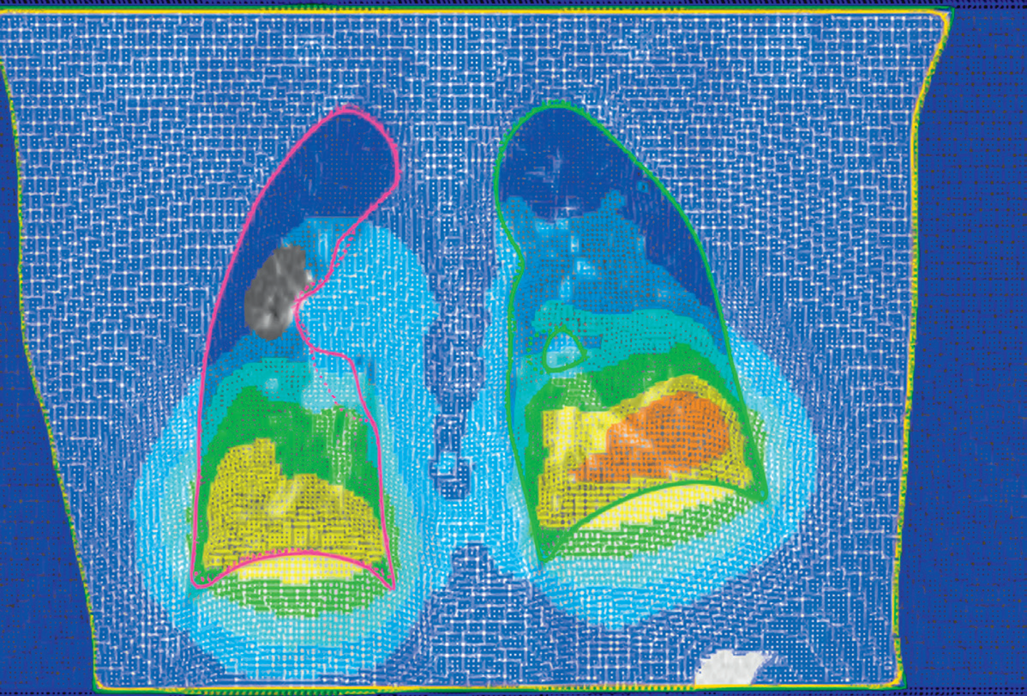
Figure 2: Fusion view showing Morfeus using sliding interfaces (left) and fixed interfaces (right) for a thorax case where the lung was used as driving force.Figure 3: Side-by-side fusion view of ANACONDA (left) and rigid registration (right) between planning CT and CBCT for a head and neck case. ROIs are indicated with solid lines (planning CT) and dashed lines (CBCT).
USING DIR
Contouring
DIR can be used to propagate structures defined in one image set to another, or to contour on fused image sets. Atlas-based and modelbased segmentation can also be used, along with all conventional contouring tools.
Dose deformation
DIR can be used to map any dose calculated in RayStation, or imported from another system, to another image set. DICOM export of deformed doses is supported.
Dose tracking
After calculating dose in several cone-beam CT datasets, the doses can be deformed back to the diagnostic-scan-based treatment plan and accumulated for DVH and metrics-based analysis. These results can be extrapolated out to the last intended fraction to help clinicians determine whether the original treatment goals will be achieved.
Adaptive planning
Should the decision be made to adjust the treatment plan based on the results from te dose tracking module in RayStation, the clinician can launch an adaptive planning tool that automates the IMRT process and compensates for dose coverage issues or adjusted goals. More information about adaptive planning and dose tracking can be found in the RaySearch white paper “Adaptive Radiation Therapy using RayStation”.
Figure 4: Deformed grid visualization (ANACONDA) showing deformation between inhalation and exhalation phases for a thorax case.
REFERENCES
1) Kadoya K, et al. Multi-institutional validation study of commercially available deformable image registration software for thoracic images. To appear in: International Journal of Radiation Oncology\*Biology\*Physics.
2) Brock, KK, et al. Accuracy of finite element model-based multi-organ deformable image registration, Medical Physics, 32(6):1647-1659, 2005.
3) Velec M, et al. (2015). Evaluation of Biomechanical Deformable Image Registration (DIR) in a Commercial Radiation Therapy Planning System. International Journal of Radiation Oncology\*Biology\*Physics, 93(3), S215–S216. http://doi.org/10.1016/j.ijrobp.2015.07.519
4) Logg A, et al. Automated Solution of Differential Equations by the Finite Element Method. Springer, 2012.
5) NETGEN: http://www.hpfem.jku.at/netgen/
6) Weistrand O, Svensson S. The ANACONDA algorithm for deformable image registration in radiotherapy. Medical Physics, 42(1):40-53, 2015.
7) Vestergaard A, et al. The potential of MRI-guided online adaptive re-optimization in radiotherapy of urinary bladder cancer. Radiotherapy and Oncology, 118:154-159, 2016.