UP-TO-DATE TREATMENT, CARE AND SUPPORT
Leeds Cancer Centre opened in 2008 and is part of Leeds Teaching Hospitals NHS Trust, which has more than 18,000 employees. More than 7,500 new patients are treated annually from a catchment population of around 2.8 million people. Specialists at Leeds Cancer Centre diagnose and treat cancer for the people of the city and the nearby Yorkshire region, providing some of the most up-to-date treatment, care and support for cancer patients.
Radiation therapy treatments available include external beam radiotherapy, brachytherapy and gamma knife. A total of ten clinical linear accelerators (linacs) are installed at the center, along with two dedicated research-funded linacs with advanced image-guided radiation therapy capabilities.
A major equipment refresh program will see ten new Elekta Versa HD and three Phillips CT-Sims installed by the end of 2020. The center also uses state-of-the-art imaging, treatment planning and treatment equipment and is committed to refreshing and updating the technology regularly.
*Subject to regulatory clearance in some markets.
ACCURATE CONTOURING
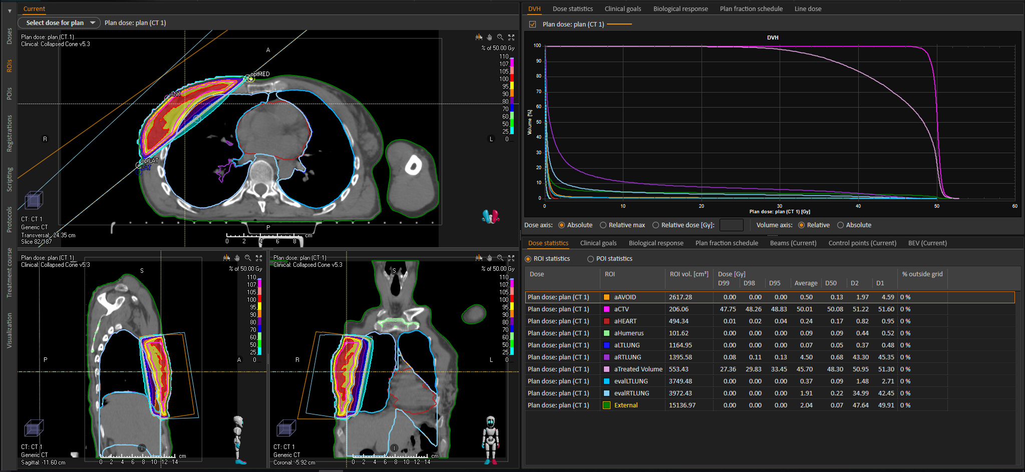
During breast radiation therapy planning, accurate lung contouring is crucial for estimating the organs at risk dose. Previously, automatic lung contours produced by the AutoBreast method in RayStation typically required manual editing to improve accuracy. Clinicians at Leeds Cancer Centre found that RaySearch thorax DL-AC model provides fast and excellent performance for ipsilateral lung contouring in the hypofractionated breast radiation therapy scenario. Given the positive results from RaySearch thorax DL-AC model, radiation oncologists in Leeds are now working closely with RaySearch to develop other machine-learning models for the safe and effective clinical implementation at other treatment sites.
We investigated the geometric and dosimetric accuracy of our new RaySearch thorax deep-learning auto-contouring (DL-AC) solution by comparing them to both reference (manual) contours and contours from the AutoBreast method for a cohort of 10 cancer patients, including left and right sided breast and chest wall treatments. Comparison of the two methods indicated that DL-AC produces accurate contours and V8Gy values, which closely approximate the reference values. AutoBreast generally overpredicts lung volume relative to reference, with less geometric and absolute dosimetric performance. However, for relative DVH statistics, error cancellation renders the overall result close to that obtained with both reference and DL-AC lung contours.
SCRIPT-DRIVEN QUALITY MANAGEMENT WORKFLOW
The team at Leeds Cancer Centre is now developing a script-driven quality management workflow for both DL-AC model training on local data and semi-automated evaluation. This approach will provide clinical confidence in models produced locally from clinical data. Assessment of clinical confidence and utility in RayStation deep learning technologies in Leeds hinges on assessment of the following criteria:
- Geometric accuracy
- Dosimetric impact
- Efficiency
- Independent, automated checking of patient specific DL-AC output
As a result of this comprehensive approach, coupled with the quality of the DL-AC contours produced by the RaySearch model, clinicians in Leeds were able to implement DL-AC derived organs at risk structures for clinical use.
A RETROSPECTIVE STUDY
10 retrospective breast patient plans were used for this analysis. These cases all contained AutoBreast lung contours from planning and had 40Gy in 15 SMLC FFF plans, which was standard practice at Leeds Cancer Centre. Further ipsilateral lung contours were generated by two methods in RayStation. Firstly, the RaySearch thorax DL-AC model was run to produce left and right lung contours. Secondly, reference ipsilateral lung contours were produced by use of the 3D-region growing tool and manual editing.
Threshold values were adjusted to fully contour lung and exclude as much airway as possible. Holes and small contours (<1cc) were removed using the standard tool. Manual editing was performed on a lung window, to exclude diaphragm and airways, and to include any lung not contained in the grown region and correct any other visually detected errors.
Dice similarity coefficient (DSC) was computed for AutoBreast and RaySearch thorax DL-AC lung contours, relative to the reference. DSC was also calculated for the AutoBreast and RaySearch thorax DL-AC model lung intersections with V25% ROI, again with the intersection volume of the manual delineation as reference. Total volumes and V25% overlap volumes were compared for all three sets of lung contours. Absolute (cc) and relative (%) V12.0Gy (30%) statistics were calculated on the 40Gy plan (consistent with V8Gy on a 26Gy plan). Lung contours were visually evaluated by an experienced dosimetrist.
SMALLER MEAN ABSOLUTE ERRORS
On all metrics, the RaySearch thorax DL-AC model showed smaller mean absolute errors relative to the reference, with a smaller range than AutoBreast.
AutoBreast appears to consistently overpredict lung volume, whereas the RaySearch thorax DL-AC model shows a volume difference 95% confidence interval containing zero. DSC is generally high, >0.9, which is expected for large and relatively simple structures such as lung.